01 · For medical teams · Training model
Ureteroscopy training kidney.
A life-size adult right kidney, from the ureter to 8 minor calyces, for hands-on flexible ureteroscopy and stone retrieval training. Made in our UK workshops.
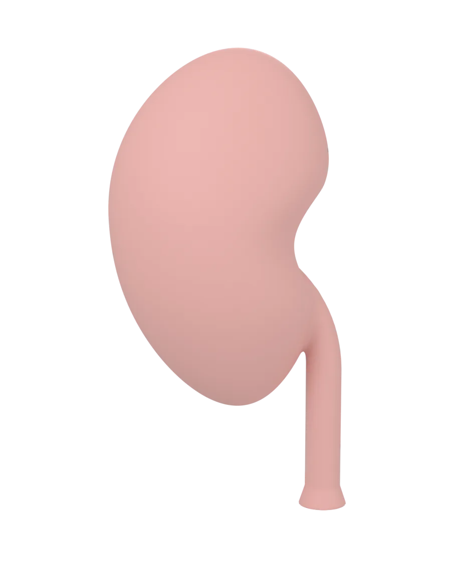
- Life-size
- Right kidney
- 8 minor calyces
- 4 lower-pole cartridges
- 14 stones
- Port for a 12/14 Fr sheath
02 · The model
Opens in two. Swaps its lower pole.
Two halves close on four pegs around a hollow collecting system: ureter, ureteropelvic junction, renal pelvis, infundibula and 8 minor calyces. A kit holds up to four cartridges, each a pair of halves with a different lower pole.
-
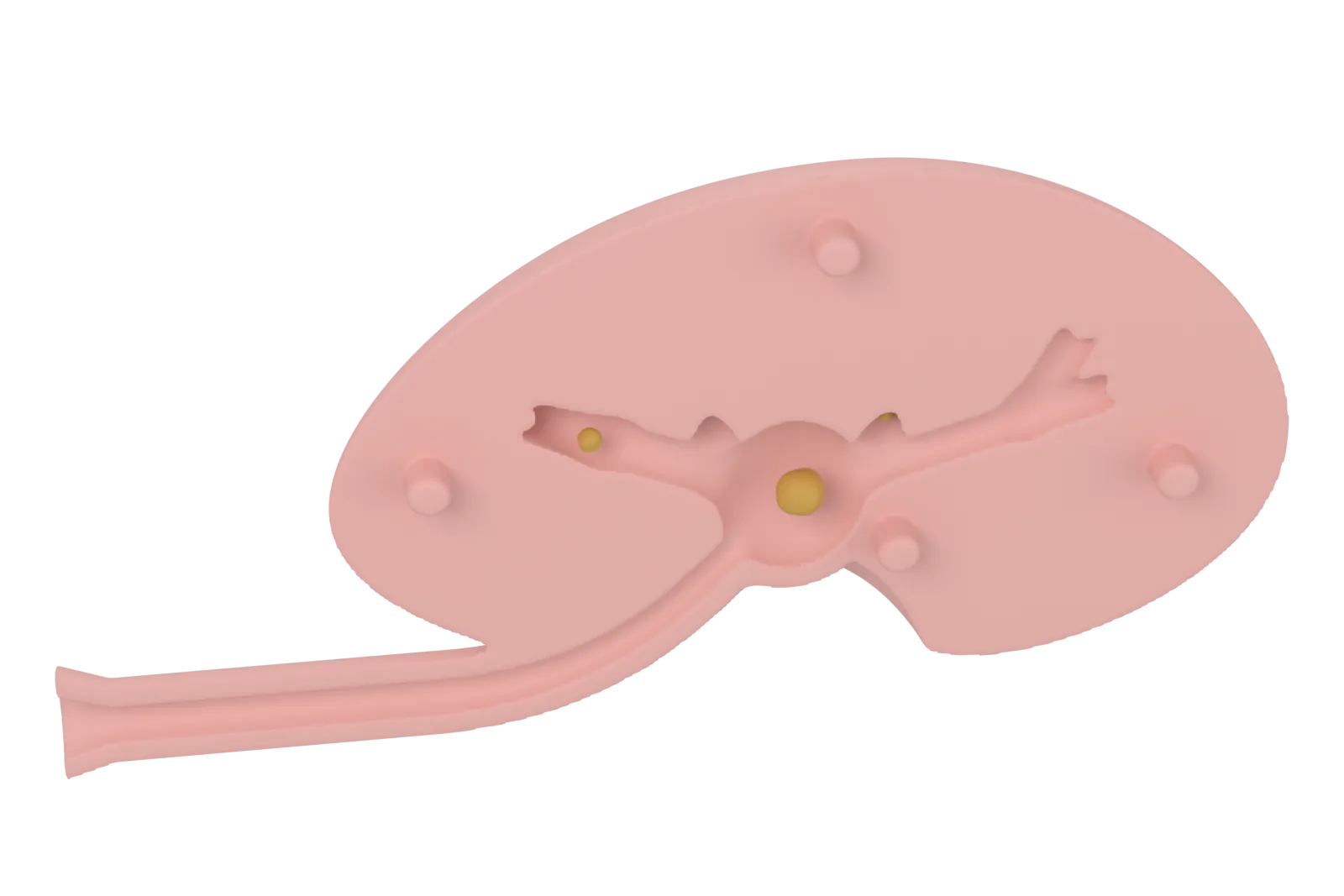
Plate 01Render Open it, place the stones, close it
Drop stones into the open posterior half, press the anterior half onto its pegs, and set the kidney in its cradle. Open it again afterwards to trace the route the scope took.
-
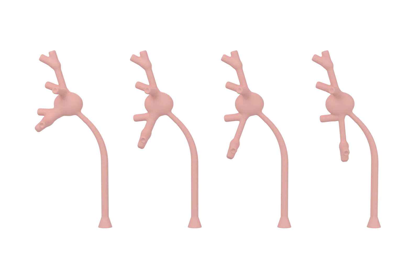
Plate 02Render Four lower poles
Favourable, standard, challenging and steep. Everything above the lower pole is the same in each, so a trainee can work the same kidney with a harder lower pole each time.
-
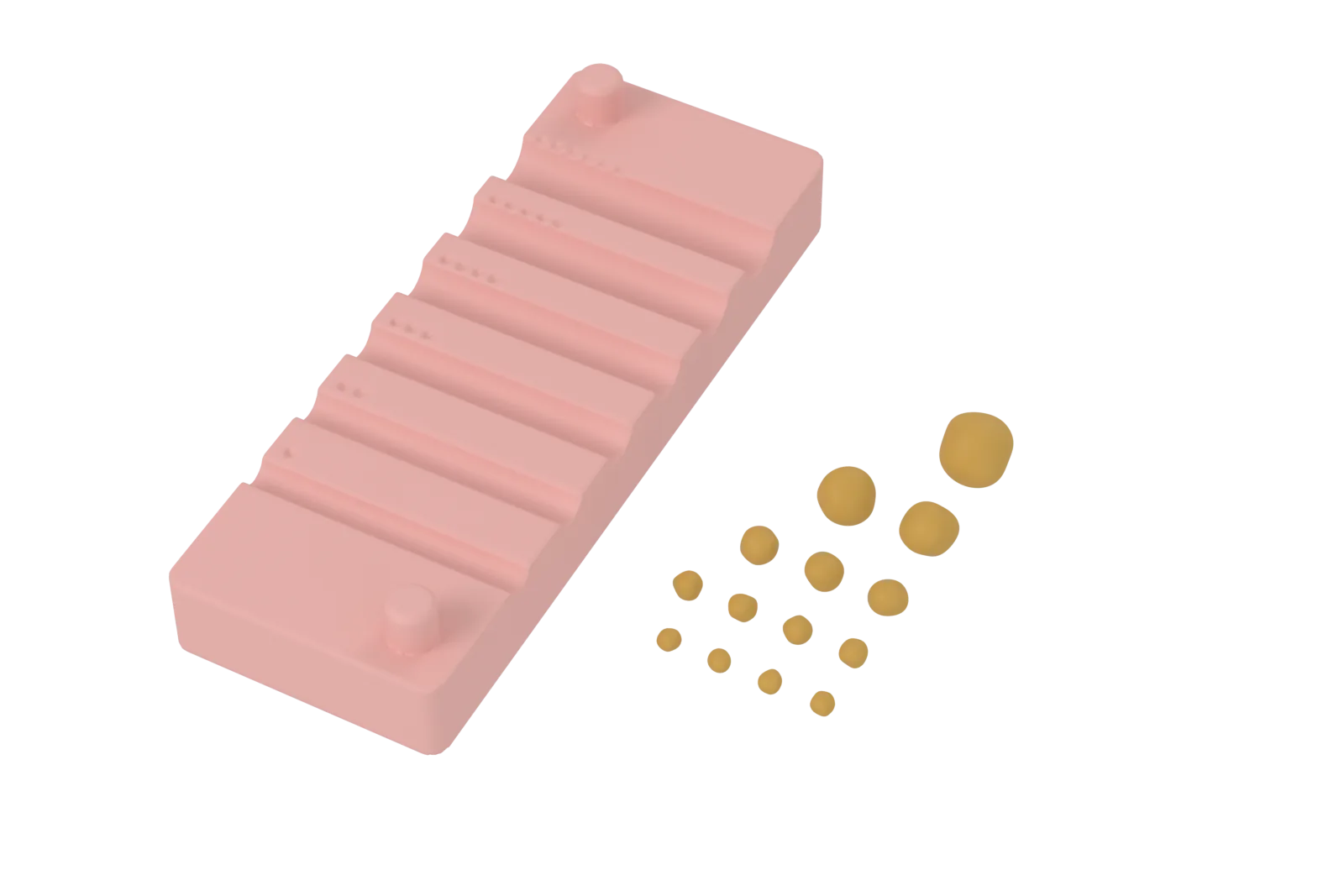
Plate 03Render Stones and a fit gauge
14 printed stones from 2.5 to 8 mm, sized to the published limits of what a basket can pull through a sheath. The fit gauge has a groove for each channel size, so you can check your scope and sheath before you start.
03 · Specification
Specification.
| Size | Shared on enquiry Two halves on four pegs, in a cradle. |
|---|---|
| Kidney | Adult right kidney, life-size. The outer form is fitted to CT scans of 33 adult right kidneys. |
| Collecting system | 8 minor calyces in a Sampaio type A1 pattern, with renal pelvis, ureteropelvic junction and proximal ureter. Hollow throughout. |
| Lower pole | Four cartridges: favourable, standard, challenging and steep. |
| Access | A port for a 12/14 Fr access sheath. The sheath stops below the ureteropelvic junction, and the scope goes on into the pelvis. |
| Stones | 14 printed stones: 4 at 2.5 mm, 4 at 3 mm, 3 at 4 mm, 2 at 6 mm and 1 at 8 mm. |
| Holds in | A cradle that turns the kidney about its long axis, medial border up, as in a patient lying on their back. |
| Material | PETG, matte light pink. Stones in amber. |
Blurred values are shared on enquiry.
04 · Measurements
Built to published anatomy.
The outer form is fitted to the median shape of 33 adult right kidneys on CT, scaled to a published mean length. The collecting system follows CT urography and endocast studies. Where studies disagree, the model takes the median of their central values.
| Measure | Model | Published adult value |
|---|---|---|
| Kidney length, pole to pole | Shared on enquiry | Shared on enquiry |
| Renal pelvis, side to side | Shared on enquiry | Shared on enquiry |
| Renal pelvis, front to back | Shared on enquiry | Shared on enquiry |
| Renal pelvis, top to bottom | Shared on enquiry | Shared on enquiry |
| Ureteropelvic junction lumen | Shared on enquiry | Shared on enquiry |
| Upper infundibulum length | Shared on enquiry | Shared on enquiry |
| Upper infundibulum width | Shared on enquiry | Shared on enquiry |
| Minor calyx cup width | Shared on enquiry | Shared on enquiry |
| Minor calyx cup length | Shared on enquiry | Shared on enquiry |
Blurred values are shared on enquiry.
The calyx necks in the mid kidney are wider than the published mean, so the mid calyces are easier to enter than in an average kidney. No published measurements of the papillae were found, so their domes are sized for printing.
This compares the design with published averages. It is not a clinical validation. Each cartridge shows one normal anatomy, and real kidneys vary widely. The renal vessels are not modelled.
05 · Scope reach
Four lower poles.
The lower pole is where flexible ureteroscopy is hardest. Each cartridge sets its lower-pole angle, infundibular length and infundibular width to a published threshold or average.
| Cartridge | Lower pole | Modelled scope reach |
|---|---|---|
| Favourable | Meets the favourable criteria of Elbahnasy et al. (1998): a wide angle and a short, wide infundibulum. | Reaches the lower calyx. |
| Standard | The median of published cohort averages. | Reaches the lower calyx. |
| Challenging | Two factors linked to lower stone-free rates: a long infundibulum and a narrow one. | Reaches the lower calyx, with less room to bend. |
| Steep | An angle that predicts failure in published series. | Out of reach by design: for practising stone repositioning, or the switch to PCNL. |
Reach is modelled for an 8.6 Fr flexible ureteroscope at its measured bend, in one plane, through rigid walls: a best case. A real collecting system distends under irrigation and gives way to the scope, so the print is harder than a patient. No scope has been through a printed model yet, so check tight fits with your own scope.
The angles are true angles in the kidney's own plane. Published angles were measured on X-ray images, where a kidney turned like this one reads lower.
06 · Training
Training with it.
For urology trainees and their trainers, and for teams who demonstrate scopes, sheaths and baskets.
-
Scope handling
- Pass from the access sheath through the ureteropelvic junction into the renal pelvis.
- Combine deflection, rotation, advance and withdrawal to enter each calyx.
- Survey all 8 calyces in a set order and return to the pelvis.
- Work the same kidney with a harder lower pole each time, from favourable to challenging.
-
Stones
- Basket a stone and pull it out through the sheath.
- Learn which stones pass: 2.5 mm through every sheath from 10/12 Fr up, 3 mm through 12/14 Fr and larger, and 4 mm through none of the sheaths tested (Randall et al., 2021).
- Move a stone out of a calyx the scope cannot reach, into one it can.
- Plan for the 6 and 8 mm stones, which need repositioning or lithotripsy.
-
Anatomy
- Name the upper, mid and lower calyx groups, and the front and back rows.
- Relate the lower-pole angle, infundibular length and width to the chance of clearing a stone.
-
What it does not show
- The walls are rigid and dry. A real collecting system distends under irrigation and gives way to the scope.
- Printed stones do not break up, so they suit basketing, not laser work.
- There is no mucosa, bleeding or renal vessels, and each cartridge is one anatomy.
07 · Use and care
Use and care.
- For ureteroscopy training and product demonstration only. It is not a medical device or a clinical tool, and it is not for use with patients.
- Use only clean scopes, sheaths and baskets that have not been used on patients.
- Use it dry. It is not sealed, so do not run irrigation through it.
- Wipe it with 70% isopropyl alcohol and let it dry. Do not soak it, and try any other disinfectant wipe on a hidden spot first.
- Do not leave it on a car dashboard in the sun. Do not sterilise or autoclave it, or put it in a dishwasher.
08 · Start a brief
Ask about the kidney.
Tell us your team, how many kits and which cartridges you need, the scopes and sheaths you use, and when.