Bronchoscopy training airway.
A life-size adult airway, from the trachea to 36 subsegmental openings, for hands-on bronchoscopy training. Made in our UK workshops.
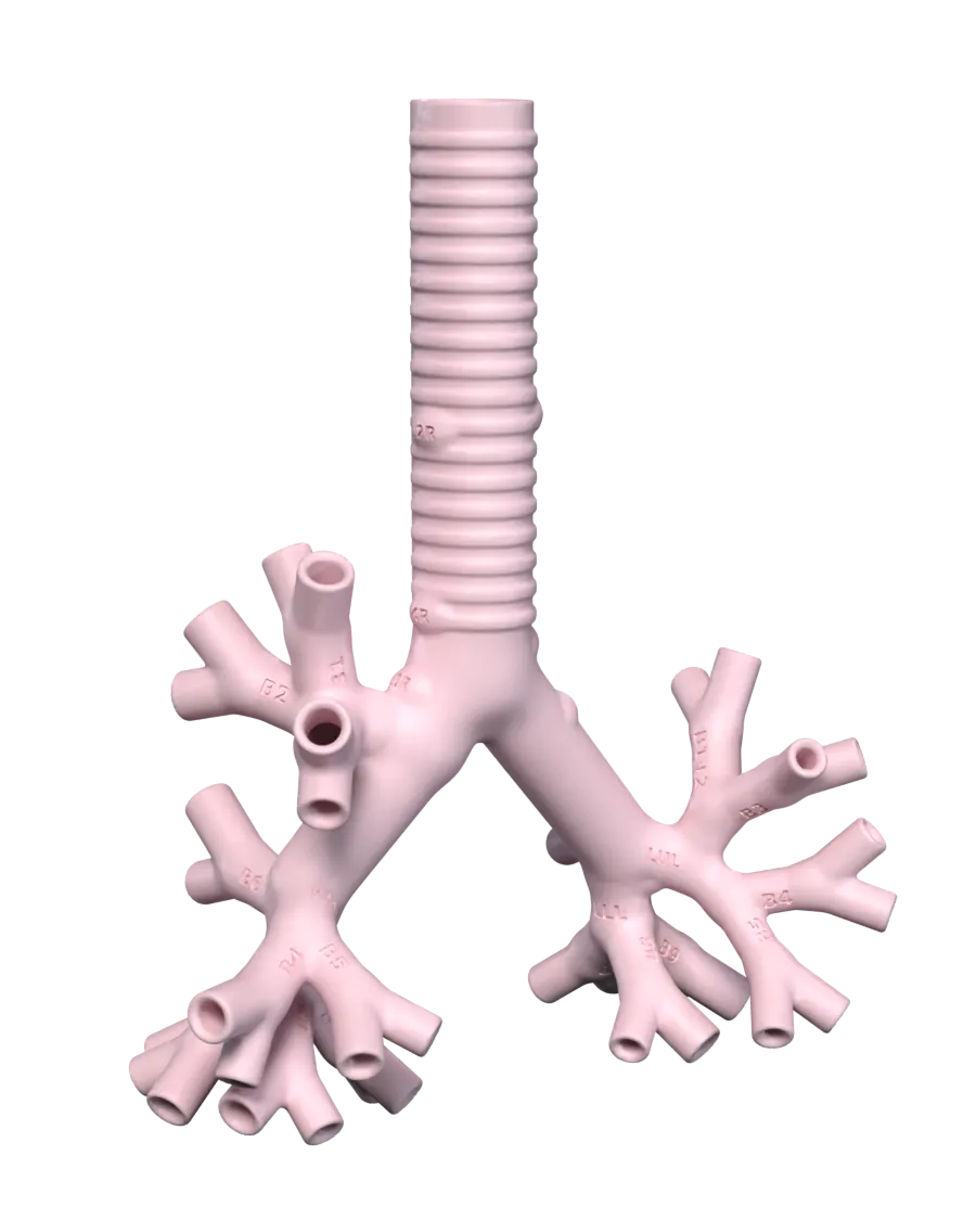
One piece. Hollow. Labelled.
A hollow airway in one piece, from the cricoid level of the trachea down to the subsegmental bronchi. It follows the standard adult branching pattern, and every opening has a rounded rim.
-
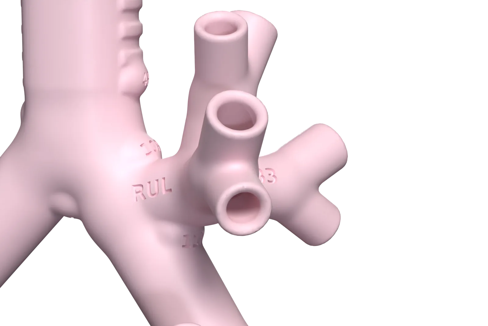
Every lobe and segment, named
23 labels are engraved into the surface: 5 lobes and 18 segments. They read upright with the trachea at the top, the way a scope goes in.
-
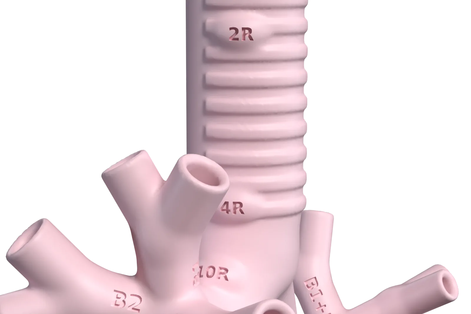
Nine lymph-node stations
2R, 2L, 4R, 4L, 7, 10R, 10L, 11R and 11L, named to the IASLC map. Each is a low dome in the airway wall with its name engraved. Positions are approximate: several IASLC borders are set by blood vessels the model does not include.
-
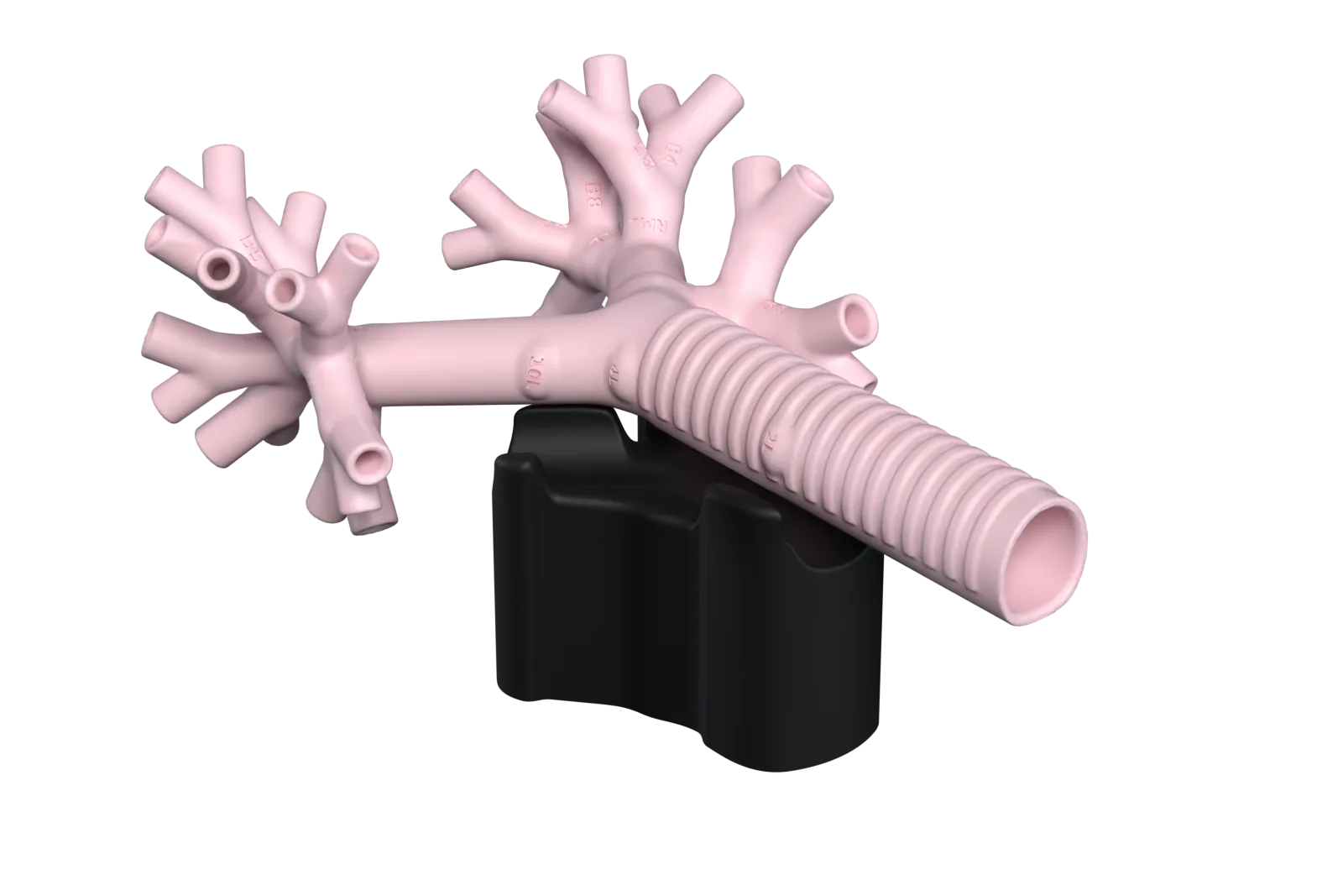
Holds still in its cradle
The airway lies on its back in a drop-in cradle and lifts straight out. Stations 4R, 4L, 7, 10R and 10L stay in view.
Specification.
-
Size
141 × 74 × 185 mm, one piece.
-
Wall
1.9 mm, hollow throughout. Open at the trachea and at 36 distal openings.
-
Trachea
17.1 mm side to side, 19.0 mm front to back. D-shaped, with a flat posterior wall and 18 cartilage rings on the outside.
-
Bronchi
5 lobar and 18 segmental bronchi, 36 subsegmental openings. Each subsegmental tube is at least 16 mm long, so a scope can be parked in it.
-
Narrowest airway
4.0 mm.
-
Material
PLA.
Built to adult measurements.
The model is built to the average airway of adult men in a CT study of 1,143 men (Mi et al., PLoS ONE, 2015). Measured on the design, each central dimension is within half a standard deviation of the published mean, and each branching angle within about a fifth.
| Measure | Model | Adult male mean |
|---|---|---|
| Trachea, side to side | 17.1 mm | 17.1 ± 2.6 mm |
| Trachea, front to back | 19.0 mm | 19.0 ± 2.3 mm |
| Trachea length, cricoid to carina | 112.7 mm | 107.8 ± 13.2 mm |
| Right main bronchus diameter | 14.1 mm | 14.1 ± 1.9 mm |
| Right main bronchus length | 16.0 mm | 14.1 ± 4.5 mm |
| Left main bronchus diameter | 12.8 mm | 13.1 ± 1.7 mm |
| Left main bronchus length | 51.7 mm | 50.0 ± 6.3 mm |
| Right upper lobe bronchus diameter | 9.9 mm | 9.9 ± 1.8 mm |
| Bronchus intermedius diameter | 12.1 mm | 12.9 ± 1.8 mm |
| Bronchus intermedius length | 30.2 mm | 32.3 ± 6.7 mm |
Further out, the segmental bronchi measure 4.0–7.4 mm and the 36 openings 4.0–6.4 mm. They follow published branch-by-branch ratios (Florens et al., 2011), with a 4.0 mm floor so an ultrathin scope can always pass. The lobar angles follow Kahraman et al. (2023).
This compares the design with published averages. It is not a clinical validation. The model shows one normal anatomy, without variants such as a tracheal bronchus. Women's central airways are on average 13–21% narrower.
Which scopes reach where.
No airway is narrower than 4.0 mm. Every scope up to 6.2 mm enters all five lobar bronchi.
| Scope outer diameter | Segmental bronchi | Subsegmental openings |
|---|---|---|
| Up to 4.0 mm | 18 of 18 | 36 of 36 |
| 4.9 mm | 15 of 18 | 15 of 36 |
| 5.5 mm | 13 of 18 | 7 of 36 |
| 5.8 mm | 11 of 18 | 5 of 36 |
| 6.2 mm | 8 of 18 | 3 of 36 |
| 6.6 mm | 6 of 18 | None |
An airway counts when it is at least as wide as the scope, so a 4.0 mm scope has no clearance in the narrowest ones. This compares published scope diameters with the design. Bends and friction reduce reach, and no scope has been through a finished model yet, so check tight fits with your own scope.
Training with it.
For registrars and fellows in respiratory medicine, anaesthetics, intensive care and paediatrics, and their trainers.
-
Scope handling
- Orientate the view from the main carina and the flat posterior wall of the trachea.
- Combine tip flexion, rotation, advance and withdrawal to enter a chosen lobar or segmental opening.
- Survey all 18 segmental bronchi in a set order and return to the carina.
- Plan a route to a named segment, call out each branch point, then follow it.
- Park the scope at a segmental opening and pass an instrument through the working channel.
-
Anatomy and scope choice
- Name the 5 lobar and 18 segmental bronchi, then check against the labels.
- Compare the sides: three lobes and a bronchus intermedius on the right; two lobes, the lingula, a combined B1+2 and no separate B7 on the left.
- Find the nine lymph-node stations, as a reference for EBUS staging.
- Choose the scope size that will reach a given segment.
-
Paediatric training
- Children have the same lobes and segments, so the routes and names carry over.
- The model is adult-sized, so it cannot teach the tight fit of a small airway.
-
What it does not show
- The airways end at the subsegmental level, so it does not reach the peripheral lung where many nodules lie.
- There is no tissue, lesion or mucosa. The stations are markers, with no ultrasound image behind them.
- The cartilage rings are on the outside, so the inside of the trachea looks smooth through a scope.
Use and care.
- For bronchoscopy training and product demonstration only. It is not a medical device or a clinical tool, and it is not for use with patients.
- Use only clean scopes and instruments that have not been used on patients.
- If a scope drags, use a little water-based lubricant on the insertion tube. It rinses out.
- Wipe inside and out with mild soap and lukewarm water, rinse, and let it dry fully. No bleach or solvents.
- PLA starts to soften at about 55°C. Do not sterilise or autoclave it, or put it in a dishwasher.
- Carry it by the trachea and main bronchi, not by the thin outer branches.
Ask about the airway.
Tell us your team, how many airways you need, the scopes you train with, and when.